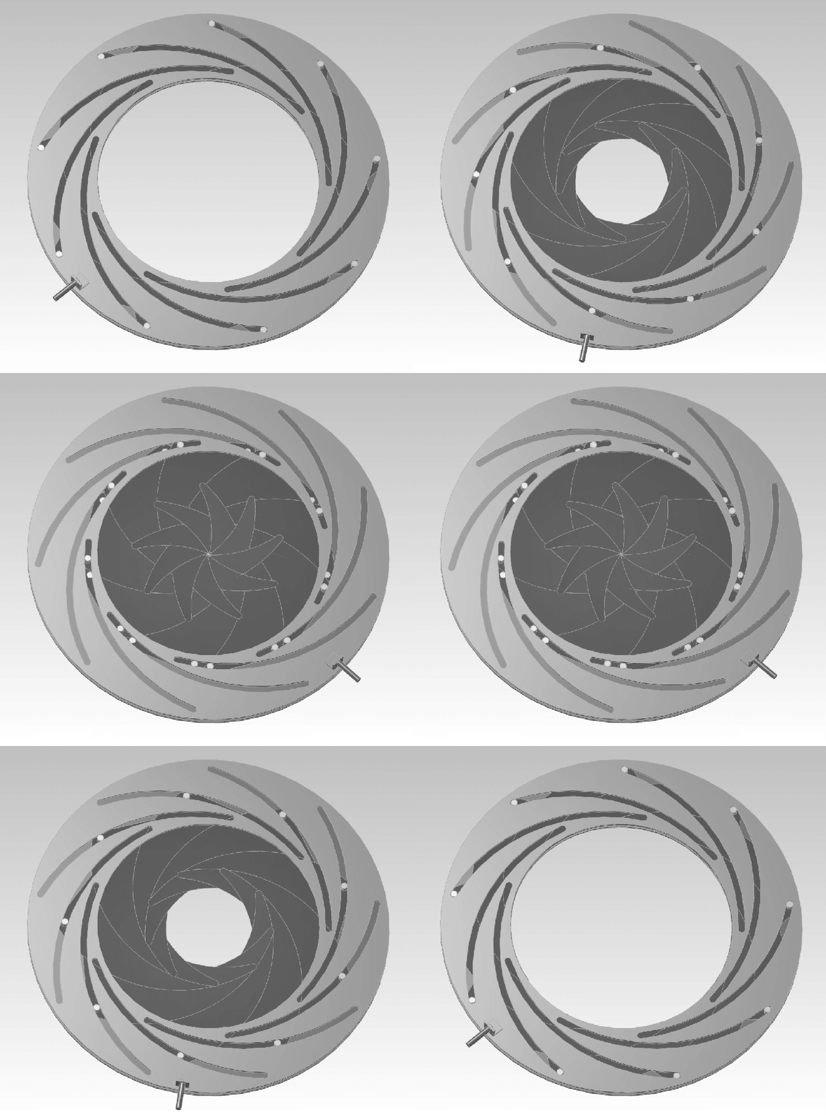
Microscopes are invaluable tools in the world of science and research, enabling us to explore the intricate details of objects and specimens at a microscopic level. At the heart of a microscope’s optical system lies a simple yet crucial component called the iris diaphragm. What is the function of an iris diaphragm, and why is it important? Keep reading to learn more!
What is an Iris Diaphragm?
An iris diaphragm, often referred to as an iris aperture or simply an iris, is a device found within the optical system of a microscope. It consists of a series of thin, overlapping metal blades arranged in a circular pattern. These blades can be adjusted to open or close the aperture, controlling the amount of light that enters the microscope’s optical path. An example would be the Thorlabs SM1D12D.

Functions of the Iris Diaphragm in a Microscope
The iris diaphragm in a microscope serves several critical functions, all geared towards optimizing the quality of the microscope’s observations:
Control of Light Intensity: The primary role of the iris diaphragm is to regulate the amount of light that illuminates the specimen under examination. By adjusting the size of the aperture, users can increase or decrease the intensity of the light. This control is essential when dealing with specimens of varying transparency or when preventing overexposure.
Depth of Field Control: The iris diaphragm also plays a significant role in controlling the depth of field in microscopy. Depth of field refers to the thickness of the specimen that appears in focus at a given time. A smaller aperture (larger f-number) increases the depth of field, making more of the specimen appear sharp at once. Conversely, a larger aperture (smaller f-number) reduces the depth of field, which can help isolate specific features or layers within the specimen.
Improving Contrast: Another benefit of adjusting the iris diaphragm is the enhancement of image contrast. By reducing the aperture size, peripheral light that enters the objective lens is minimized, reducing glare and increasing contrast. This is particularly useful when observing translucent or low-contrast specimens.

Minimizing Aberrations: In some cases, the iris diaphragm can help minimize optical aberrations that can degrade the quality of the microscope image. Aberrations like spherical or chromatic aberration can be reduced by stopping down the aperture by using a smaller iris opening to improve image clarity.
Preventing Specimen Damage: For extremely sensitive specimens, such as live cells or delicate biological samples, the iris diaphragm can limit the amount of light exposure. This precaution prevents specimen damage or photobleaching, which is the loss of fluorescence signal due to excessive light exposure.
In conclusion, the iris diaphragm in a microscope is a versatile and indispensable tool for fine-tuning the quality of illumination and image formation. It allows researchers and microscopists to adjust lighting conditions and image characteristics to obtain the best possible observations of specimens. Whether it’s controlling light intensity, depth of field, contrast, or minimizing aberrations, the iris diaphragm empowers scientists to unlock the hidden beauty of the microcosm.